publications
publications by categories in reversed chronological order. generated by jekyll-scholar.
Updated on 08/2025. Please cross-check with my Google Scholar or ResearchGate profiles.
2025
- BOE
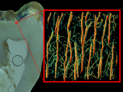
 Label-free visualisation of histological features in human teeth using autofluorescence imagingSeunghwan Goldmund Lee, Elsa Vennat, Kwangseuk Kyhm, and 1 more authorBiomedical Optics Express, 2025
We show that autofluorescence imaging can be used as a label-free modality to visualise histological features of human teeth. The autofluorescence emitted by enamel, dentin, predentin, and interglobular dentin (IGD) allows observing structural details that would otherwise require complex staining or even complementary electron microscopy or X-ray imaging. The simultaneous acquisition of autofluorescence and conventional staining fluorescence images by confocal microscope also provides important advantages to visualise occluded dentin porosity or the presence of porosity within IGD. However, autofluorescence imaging is practically limited by photobleaching. We, therefore, quantified the photobleaching extent in 3D and showed that, in confocal microscopy, the whole illuminated volume is bleached during acquisition. To mitigate this phenomenon, we analysed the photobleaching behaviour in enamel, dentin, predentin, and IGD as a function of laser power. We found that the measured decay rate cannot be modelled by a single population decay of fluorophores, highlighting the fact that autofluorescence probably arises from multiple sources. This analysis also allowed defining a laser excitation power where the photobleaching effect saturates.
@article{lee2025label, title = {Label-free visualisation of histological features in human teeth using autofluorescence imaging}, author = {Lee, Seunghwan Goldmund and Vennat, Elsa and Kyhm, Kwangseuk and Gourrier, Aur{\'e}lien}, journal = {Biomedical Optics Express}, volume = {16}, number = {7}, pages = {2792--2807}, year = {2025}, publisher = {Optica Publishing Group}, url = {https://doi.org/10.1364/BOE.564484}, doi = {10.1364/BOE.564484}, } - PLOSOne
 Cellular porosity in dentin exhibits complex network characteristics with spatio-temporal fluctuationsLucas Chatelain, Nicolas Tremblay, Elsa Vennat, and 3 more authorsPLoS One, 2025
According to the current hydrodynamic theory, teeth sensitivity is mediated by odontoblast cell processes which can be activated by fluid flow in the pericellular space of bulk dentin. To better understand the possible spatial extent of such phenomena, we investigated the topology and connectivity of dentinal porosity of a healthy human tooth. Using confocal fluorescence microscopy, we modeled the porosity as a spatial graph with edges representing dentinal tubules or lateral branches and nodes defining their connections. A large fraction of porosity channels in crown dentin was found to be interconnected, with 47% of nodes linked in a single component over a millimetric distance from the dentin-enamel junction (DEJ). However, significant differences in network topology were also observed. A sharp transition in connectivity from 83% to 43% occurred at 300 µm from the DEJ, which corresponds to an early stage of tooth formation. This was reflected in all graph metrics investigated, in particular the network resilience which dropped by a factor 2. To test the robustness of our observations, an in-depth analysis of potential remaining biases of the graph extraction was conducted. Most graph metrics considered were found to be within a 10% precision range from a manually annotated ground truth. However, path metrics, which characterize transport properties, proved very sensitive to network defects. Residual errors were classified in 4 topological classes related to fluorescence staining and confocal detection efficiency, instrumental resolution and image processing. Their relative importance was estimated using statistical and physical graph attack simulations in a broad experimental range. Our modeling thus provides a practical framework to estimate the interpretability of calculated graph metrics for a given experimental microscopy setup and image processing pipeline. Overall, this study shows that dentin porosity exhibits typical characteristics of a complex network and quantitatively emphasize the importance of the smallest lateral branches. Our results could be used to model fluid flow more accurately in order to better understand mechanosensing by odontoblasts in dentin.
@article{chatelain2025cellular,
  title = {Cellular porosity in dentin exhibits complex network characteristics with spatio-temporal fluctuations},
  author = {Chatelain, Lucas and Tremblay, Nicolas and Vennat, Elsa and Dursun, Elisabeth and Rousseau, David and Gourrier, Aur{\'e}lien},
  journal = {PLoS One},
  volume = {20},
  number = {7},
  pages = {e0327030},
  year = {2025},
  publisher = {Public Library of Science San Francisco, CA USA},
  url = {https://doi.org/10.1371/journal.pone.0327030},
  doi = {10.1371/journal.pone.0327030},
}
2024
- JSBTooth acellular extrinsic fibre cementum incremental lines in humans are formed by parallel branched Sharpey’s fibres and not by its mineral phaseLourdes R Couoh, Lauro Bucio, José Luis Ruvalcaba, and 4 more authorsJournal of Structural Biology, 2024
In humans, the growth pattern of the acellular extrinsic fibre cementum (AEFC) has been useful to estimate the age-at-death. However, the structural organization behind such a pattern remains poorly understood. In this study tooth cementum from seven individuals from a Mexican modern skeletal series were analyzed with the aim of unveiling the AEFC collagenous and mineral structure using multimodal imaging approaches. The organization of collagen fibres was first determined using: light microscopy, transmission electron microscopy (TEM), electron tomography, and plasma FIB scanning electron microscopy (PFIB-SEM) tomography. The mineral properties were then investigated using: synchrotron small-angle X-ray scattering (SAXS) for T-parameter (correlation length between mineral particles); synchrotron X-ray diffraction (XRD) for L-parameter (mineral crystalline domain size estimation), alignment parameter (crystals preferred orientation) and lattice parameters a and c; as well as synchrotron X-ray fluorescence for spatial distribution of calcium, phosphorus and zinc. Results show that Sharpey’s fibres branched out fibres that cover and uncover other collagen bundles forming aligned arched structures that are joined by these same fibres but in a parallel fashion. The parallel fibres are not set as a continuum on the same plane and when they are superimposed project the AEFC incremental lines due to the collagen birefringence. The orientation of the apatite crystallites is subject to the arrangement of the collagen fibres, and the obtained parameter values along with the elemental distribution maps, revealed this mineral tissue as relatively homogeneous. Therefore, no intrinsic characteristics of the mineral phase could be associated with the alternating AEFC incremental pattern.
@article{couoh2024tooth,
  title = {Tooth acellular extrinsic fibre cementum incremental lines in humans are formed by parallel branched Sharpey’s fibres and not by its mineral phase},
  author = {Couoh, Lourdes R and Bucio, Lauro and Ruvalcaba, Jos{\'e} Luis and Manoel, Britta and Tang, Tengteng and Gourrier, Aur{\'e}lien and Grandfield, Kathryn},
  journal = {Journal of Structural Biology},
  volume = {216},
  number = {2},
  pages = {108084},
  year = {2024},
  publisher = {Elsevier},
  url = {https://doi.org/10.1016/j.jsb.2024.108084},
  doi = {10.1016/j.jsb.2024.108084}
}
2023
- ActaBiomaterThe elasto-plastic nano-and microscale compressive behaviour of rehydrated mineralised collagen fibresAlexander Groetsch, Aurélien Gourrier, Daniele Casari, and 5 more authorsActa biomaterialia, 2023
The hierarchical design of bio-based nanostructured materials such as bone enables them to combine unique structure-mechanical properties. As one of its main components, water plays an important role in bone’s material multiscale mechanical interplay. However, its influence has not been quantified at the length-scale of a mineralised collagen fibre. Here, we couple in situ micropillar compression, and simultaneous synchrotron small angle X-ray scattering (SAXS) and X-ray diffraction (XRD) with a statistical constitutive model. Since the synchrotron data contain statistical information on the nanostructure, we establish a direct connection between experiment and model to identify the rehydrated elasto-plastic micro- and nanomechanical fibre behaviour. Rehydration led to a decrease of 65%-75% in fibre yield stress and compressive strength, and 70% in stiffness with a 3x higher effect on stresses than strains. While in agreement with bone extracellular matrix, the decrease is 1.5-3x higher compared to micro-indentation and macro-compression. Hydration influences mineral more than fibril strain with the highest difference to the macroscale when comparing mineral and tissue levels. The effect of hydration seems to be strongly mediated by ultrastructural interfaces while results provide insights towards mechanical consequences of reported water-mediated structuring of bone apatite. The missing reinforcing capacity of surrounding tissue for an excised fibril array is more pronounced in wet than dry conditions, mainly related to fibril swelling. Differences leading to higher compressive strength between mineralised tissues seem not to depend on rehydration while the lack of kink bands supports the role of water as an elastic embedding influencing energy-absorption mechanisms.
@article{groetsch2023elasto,
  title = {The elasto-plastic nano-and microscale compressive behaviour of rehydrated mineralised collagen fibres},
  author = {Groetsch, Alexander and Gourrier, Aur{\'e}lien and Casari, Daniele and Schwiedrzik, Jakob and Shephard, Jonathan D and Michler, Johann and Zysset, Philippe K and Wolfram, Uwe},
  journal = {Acta biomaterialia},
  volume = {164},
  pages = {332--345},
  year = {2023},
  publisher = {Elsevier},
  url = {https://doi.org/10.1016/j.actbio.2023.03.041},
  doi = {10.1016/j.actbio.2023.03.041}
}
2022
- ActaBiomaterBone mineral organization at the mesoscale: A review of mineral ellipsoids in bone and at bone interfacesChiara Micheletti, Ariana Hurley, Aurélien Gourrier, and 4 more authorsActa Biomaterialia, 2022
Much debate still revolves around bone architecture, especially at the nano- and microscale. Bone is a remarkable material where high strength and toughness coexist thanks to an optimized composition of mineral and protein and their hierarchical organization across several distinct length scales. At the nanoscale, mineralized collagen fibrils act as building block units. Despite their key role in biological and mechanical functions, the mechanisms of collagen mineralization and the precise arrangement of the organic and inorganic constituents in the fibrils remains not fully elucidated. Advances in three-dimensional (3D) characterization of mineralized bone tissue by focused ion beam-scanning electron microscopy (FIB-SEM) revealed mineral-rich regions geometrically approximated as prolate ellipsoids, much larger than single collagen fibrils. These structures have yet to become prominently recognized, studied, or adopted into biomechanical models of bone. However, they closely resemble the circular to elliptical features previously identified by scanning transmission electron microscopy (STEM) in two-dimensions (2D). Herein, we review the presence of mineral ellipsoids in bone as observed with electron-based imaging techniques in both 2D and 3D with particular focus on different species, anatomical locations, and in proximity to natural and synthetic biomaterial interfaces. This review reveals that mineral ellipsoids are a ubiquitous structure in all the bones and bone-implant interfaces analyzed. This largely overlooked hierarchical level is expected to bring different perspectives to our understanding of bone mineralization and mechanical properties, in turn shedding light on structure-function relationships in bone.
@article{micheletti2022bone, title = {Bone mineral organization at the mesoscale: A review of mineral ellipsoids in bone and at bone interfaces}, author = {Micheletti, Chiara and Hurley, Ariana and Gourrier, Aur{\'e}lien and Palmquist, Anders and Tang, Tengteng and Shah, Furqan A and Grandfield, Kathryn}, journal = {Acta Biomaterialia}, volume = {142}, pages = {1--13}, year = {2022}, publisher = {Elsevier}, url = {https://doi.org/10.1016/j.actbio.2022.02.024}, doi = {10.1016/j.actbio.2022.02.024} } - Autofluorescence Loss in Photobleaching for Human Dentin ex vivoSeunghwan Goldmund Lee, Minwoo Kim, Sunghee Jeong, and 5 more authorsCurrent Optics and Photonics, 2022
@article{lee2022autofluorescence,
  title = {Autofluorescence Loss in Photobleaching for Human Dentin ex vivo},
  author = {Lee, Seunghwan Goldmund and Kim, Minwoo and Jeong, Sunghee and Hwang, Jaejoon and Kim, Jisu and Gourrier, Aur{\'e}lien and Vial, Jean Claude and Kyhm, Kwangseuk},
  journal = {Current Optics and Photonics},
  volume = {6},
  number = {1},
  pages = {86--91},
  year = {2022},
  publisher = {Optical Society of Korea},
  url = {https://doi.org/10.3807/COPP.2022.6.1.086},
  doi = {10.3807/COPP.2022.6.1.086}
}
2020
- JSBEllipsoidal mesoscale mineralization pattern in human cortical bone revealed in 3D by plasma focused ion beam serial sectioningDakota M Binkley, Joseph Deering, Hui Yuan, and 2 more authorsJournal of Structural Biology, 2020
Visualizing bone mineralization and collagen fibril organization at intermediate scales between the nanometer and the hundreds of microns range, is still an important challenge. Similarly, visualizing cellular components which locally affect the tissue structure requires a precision of a few tens of nanometers at maximum while spanning several tens of micrometers. In the last decade, gallium focused ion beam (FIB) equipped with a scanning electron microscope (SEM) proved to be an extremely valuable structural tool to meet those ends. In this study, we assess the capability of a recent plasma FIB-SEM technology which provides a potential increase in measurement speed over gallium FIB-SEM, thus paving the way to larger volume analysis. Nanometer-scale layers of demineralized and mineralized unstained human femoral lamellar bone were sequentially sectioned over volumes of 6–16,000 μm3. Analysis of mineralized tissue revealed prolate ellipsoidal mineral clusters measuring approximately 1.1 µm in length by 700 nm at their maximum diameter. Those features, suggested by others in high resolution studies, appear here as a ubiquitous motif in mineralized lamellar bone over thousands of microns cubed, suggesting a heterogeneous and yet regular pattern of mineral deposition past the single collagen fibril level. This large scale view retained sufficient resolution to visualize the collagen fibrils while also partly visualizing the lacuno-canalicular network in three-dimensions. These findings are strong evidence for suitability of PFIB as a bone analysis tool and the need to revisit bone mineralization over multi-length scales with mineralized tissue.
@article{binkley2020ellipsoidal,
  title = {Ellipsoidal mesoscale mineralization pattern in human cortical bone revealed in 3D by plasma focused ion beam serial sectioning},
  author = {Binkley, Dakota M and Deering, Joseph and Yuan, Hui and Gourrier, Aur{\'e}lien and Grandfield, Kathryn},
  journal = {Journal of Structural Biology},
  volume = {212},
  number = {2},
  pages = {107615},
  year = {2020},
  publisher = {Academic Press},
  url = {https://doi.org/10.1016/j.jsb.2020.107615},
  doi = {10.1016/j.jsb.2020.107615}
}
2019
- ConfProcLabel-free THG imaging of bone tissue microstructure: effect of low gravity on the lacuno-canalicular networkRachel Genthial, Emmanuel Beaurepaire, Marie-Claire Schanne-Klein, and 6 more authorsIn Advances in Microscopic Imaging II, 2019
In bone tissue, multiscale interfaces provide the structural basis of essential bone functions and contribute to its macroscopic mechanical properties. The lacuno-canalicular network (LCN) hosting the osteocytes in the bone matrix, in particular, represents a biological signature of the mechanotransduction activity in response to external biomechanical loading. We have demonstrated that label-free third-harmonic generation (THG) microscopy reveals the structure of the LCN in 3D with submicron precision over millimetric fields of view compatible with histology and can be coupled to second-harmonic generation (SHG) signals relating to the collagen organization in the bone matrix. Taking advantage of these label-free imaging methods, we investigate the impact of microgravity on the LCN structure in mice following a 1month space flight. We show that our current lack of understanding of the extent of the LCN heterogeneity at the organ level hinders the interpretation of such investigations based on a limited number of samples and we discuss the implications for future biomedical studies.
- PLOSOneThird harmonic generation imaging and analysis of the effect of low gravity on the lacuno-canalicular network of mouse boneRachel Genthial, Maude Gerbaix, Delphine Farlay, and 4 more authorsPloS one, 2019
The lacuno-canalicular network (LCN) hosting the osteocytes in bone tissue represents a biological signature of the mechanotransduction activity in response to external biomechanical loading. Using third-harmonic generation (THG) microscopy with sub-micrometer resolution, we investigate the impact of microgravity on the 3D LCN structure in mice following space flight. A specific analytical procedure to extract the LCN characteristics from THG images is described for ex vivo studies of bone sections. The analysis conducted in different anatomical quadrants of femoral cortical bone didn’t reveal any statistical differences between the control, habitat control and flight groups, suggesting that the LCN connectivity is not affected by one month space flight. However, significant variations are systematically observed within each sample. We show that our current lack of understanding of the extent of the LCN heterogeneity at the organ level hinders the interpretation of such investigations based on a limited number of samples and we discuss the implications for future biomedical studies.
@article{genthial2019third, title = {Third harmonic generation imaging and analysis of the effect of low gravity on the lacuno-canalicular network of mouse bone}, author = {Genthial, Rachel and Gerbaix, Maude and Farlay, Delphine and Vico, Laurence and Beaurepaire, Emmanuel and D{\'e}barre, Delphine and Gourrier, Aur{\'e}lien}, journal = {PloS one}, volume = {14}, number = {1}, pages = {e0209079}, year = {2019}, publisher = {Public Library of Science}, url = {https://doi.org/10.1371/journal.pone.0209079}, doi = {10.1371/journal.pone.0209079} } - ActaBiomaterCompressive behaviour of uniaxially aligned individual mineralised collagen fibres at the micro-and nanoscaleAlexander Groetsch, Aurélien Gourrier, Jakob Schwiedrzik, and 6 more authorsActa biomaterialia, 2019
The increasing incidence of osteoporotic bone fractures makes fracture risk prediction an important clinical challenge. Computational models can be utilised to facilitate such analyses. However, they critically depend on bone’s underlying hierarchical material description. To understand bone’s irreversible behaviour at the micro- and nanoscale, we developed an in situ testing protocol that allows us to directly relate the experimental data to the mechanical behaviour of individual mineralised collagen fibres and its main constitutive phases, the mineralised collagen fibrils and the mineral nanocrystals, by combining micropillar compression of single fibres with small angle X-ray scattering (SAXS) and X-ray diffraction (XRD). Failure modes were assessed by SEM. Strain ratios in the elastic region at fibre, fibril and mineral levels were found to be approximately 22:5:2 with strain ratios at the point of compressive strength of 0.23 Æ 0.11 for fibril-to-fibre and 0.07 Æ 0.01 for mineral-to-fibre levels. Mineral-to-fibre levels showed highest strain ratios around the apparent yield point, fibril-to-fibre around apparent strength. The mineralised collagen fibrils showed a delayed mechanical response, contrary to the mineral phase, which points towards preceding deformations of mineral nanocrystals in the extrafibrillar matrix. No damage was measured at the level of the mineralised collagen fibre which indicates an incomplete separation of the mineral and collagen, and an extrafibrillar interface failure. The formation of kink bands and the gradual recruitment of fibrils upon compressive loading presumably led to localised strains. Our results from a well-controlled fibrillar architecture provide valuable input for micromechanical models and computational non-linear bone strength analyses that may provide further insights for personalised diagnosis and treatment as well as bio-inspired implants for patients with bone diseases.
@article{groetsch2019compressive, title = {Compressive behaviour of uniaxially aligned individual mineralised collagen fibres at the micro-and nanoscale}, author = {Groetsch, Alexander and Gourrier, Aur{\'e}lien and Schwiedrzik, Jakob and Sztucki, Michael and Beck, Rainer J and Shephard, Jonathan D and Michler, Johann and Zysset, Philippe K and Wolfram, Uwe}, journal = {Acta biomaterialia}, volume = {89}, pages = {313--329}, year = {2019}, publisher = {Elsevier}, url = {https://doi.org/10.1016/j.actbio.2019.03.043}, doi = {10.1016/j.actbio.2019.02.053} } - ActaBiomaterAnisotropic elastic properties of human femoral cortical bone and relationships with composition and microstructure in elderlyXiran Cai, Hélène Follet, Laura Peralta, and 8 more authorsActa biomaterialia, 2019
The strong dependence between cortical bone elasticity at the millimetre-scale (mesoscale) and cortical porosity has been evidenced by previous studies. However, bone is an anisotropic composite material made by mineral, proteins and water assembled in a hierarchical structure. Whether the variations of structural and compositional properties of bone affect the different elastic coefficients at the mesoscale is not clear. Aiming to understand the relationships between bone elastic properties and compositions and microstructure, we applied state-of-the-art experimental modalities to assess these aspects of bone characteristics. All elastic coefficients (stiffness tensor of the transverse isotropic bone material), structure of the vascular pore network, collagen and mineral properties were measured in 52 specimens from the femoral diaphysis of 26 elderly donors. Statistical analyses and micromechanical modeling showed that vascular pore volume fraction and the degree of mineralization of bone are the most important determinants of cortical bone anisotropic mesoscopic elasticity. Though significant correlations were observed between collagen properties and elasticity, their effects in bone mesoscopic elasticity were minor in our data. This work also provides a unique set of data exhibiting a range of variations of compositional and microstructural cortical bone properties in the elderly and gives strong experimental evidence and basis for further development of biomechanical models for human cortical bone.
@article{cai2019anisotropic,
  title = {Anisotropic elastic properties of human femoral cortical bone and relationships with composition and microstructure in elderly},
  author = {Cai, Xiran and Follet, H{\'e}l{\`e}ne and Peralta, Laura and Gardegaront, Marc and Farlay, Delphine and Gauthier, R{\'e}my and Yu, Boliang and Gineyts, Evelyne and Olivier, C{\'e}cile and Langer, Max and others},
  journal = {Acta biomaterialia},
  volume = {90},
  pages = {254--266},
  year = {2019},
  publisher = {Elsevier},
  url = {https://doi.org/10.1016/j.actbio.2019.03.043},
  doi = {10.1016/j.actbio.2019.03.043}
}
2018
- ActaBiomaterThe three-dimensional arrangement of the mineralized collagen fibers in elephant ivory and its relation to mechanical and optical propertiesM. Albéric, A. Gourrier, W. Wagermaier, and 2 more authorsActa Biomaterialia, 2018
Elephant tusks are composed of dentin or ivory, a hierarchical and composite biological material made of mineralized collagen fibers (MCF). The specific arrangement of the MCF is believed to be responsible for the optical and mechanical properties of the tusks. Especially the MCF organization likely contributes to the formation of the bright and dark checkerboard pattern observed on polished sections of tusks (Schreger pattern). Yet, the precise structural origin of this optical motif is still controversial. We hereby address this issue using complementary analytical methods (small and wide angle X-ray scattering, cross-polarized light microscopy and scanning electron microscopy) on elephant ivory samples and show that MCF orientation in ivory varies from the outer to the inner part of the tusk. An external cohesive layer of MCF with fiber direction perpendicular to the tusk axis wraps the mid-dentin region, where the MCF are oriented mainly along the tusk axis and arranged in a plywood-like structure with fiber orientations oscillating in a narrow angular range. This particular oscillating-plywood structure of the MCF and the birefringent properties of the collagen fibers, likely contribute to the emergence of the Schreger pattern, one of the most intriguing macroscopic optical patterns observed in mineralized tissues and of great importance for authentication issues in archeology and forensic sciences.
@article{alberic_three-dimensional_2018, title = {The three-dimensional arrangement of the mineralized collagen fibers in elephant ivory and its relation to mechanical and optical properties}, author = {Albéric, M. and Gourrier, A. and Wagermaier, W. and Fratzl, P. and Reiche, I.}, journal = {Acta Biomaterialia}, volume = {72}, pages = {342--351}, year = {2018}, publisher = {Elsevier}, url = {https://doi.org/10.1016/j.actbio.2018.02.016}, doi = {10.1016/j.actbio.2018.02.016} } - ActaBiomaterUltrafine heat-induced structural perturbations of bone mineral at the individual nanocrystal levelM. Verezhak, E.F. Rauch, M. Véron, and 4 more authorsActa Biomaterialia, 2018
The nanoscale characteristics of the mineral phase in bone tissue such as nanocrystal size, organization, structure and composition have been identified as potential markers of bone quality. However, such characterization remains challenging since it requires combining structural analysis and imaging modalities with nanoscale precision. In this paper, we report the first application of automated crystal orientation mapping using transmission electron microscopy (ACOM-TEM) to the structural analysis of bone mineral at the individual nanocrystal level. By controlling the nanocrystal growth of a cortical bovine bone model artificially heated up to 1000 °C, we highlight the potential of this technique. We thus show that the combination of sample mapping by scanning and the crystallographic information derived from the collected electron diffraction patterns provides a more rigorous analysis of the mineral nanostructure than standard TEM. In particular, we demonstrate that nanocrystal orientation maps yield valuable information for dimensional analysis. Furthermore, we show that ACOM-TEM has sufficient sensitivity to distinguish between phases with close crystal structures and we address unresolved questions regarding the existence of a hexagonal to monoclinic phase transition induced by heating. This first study therefore opens new perspectives in bone characterization at the nanoscale, a daunting challenge in the biomedical and archaeological fields, which could also prove particularly useful to study the mineral characteristics of tissue grown at the interface with biomaterials implants.
@article{verezhak_ultrafine_2018, title = {Ultrafine heat-induced structural perturbations of bone mineral at the individual nanocrystal level}, author = {Verezhak, M. and Rauch, E.F. and Véron, M. and Lancelon-Pin, C. and Putaux, J.-L. and Plazanet, M. and Gourrier, A.}, journal = {Acta Biomaterialia}, volume = {73}, pages = {500--508}, year = {2018}, publisher = {Elsevier}, url = {https://doi.org/10.1016/j.actbio.2018.04.004}, doi = {10.1016/j.actbio.2018.04.004} } - PLOSOneTime-domain THz spectroscopy of the characteristics of hydroxyapatite provides a signature of heating in bone tissueMarie Plazanet, Jordanka Tasseva, Paolo Bartolini, and 7 more authorsPloS one, 2018
Because of the importance of bone in the biomedical, forensic and archaeological contexts, new investigation techniques are constantly required to better characterize bone ultrastructure. In the present paper, we provide an extended investigation of the vibrational features of bone tissue in the 0.1-3 THz frequency range by time-domain THz spectroscopy. Their assignment is supported by a combination of X-ray diffraction and DFT-normal modes calculations. We investigate the effect of heating on bone tissue and synthetic calciumphosphates compounds with close structure and composition to bone mineral, including stoichiometric and non-stoichiometric hydroxyapatite (HA), tricalcium phosphate, calcium pyrophosphate and tetracalcium phosphate. We thus demonstrate that the narrow vibrational mode at 2.1 THz in bone samples exposed to thermal treatment above 750 ˚C arises from a lattice mode of stoichiometric HA. This feature is also observed in the other synthetic compounds, although weaker or broader, but is completely smeared out in the non-stoichiometric HA, close to natural bone mineral composition, or in synthetic poorly crystalline HA powder. The THz spectral range therefore provides a clear signature of the crystalline state of the investigated bone tissue and could, therefore be used to monitor or identify structural transitions occurring in bone upon heating.
@article{plazanet2018time,
  title = {Time-domain THz spectroscopy of the characteristics of hydroxyapatite provides a signature of heating in bone tissue},
  author = {Plazanet, Marie and Tasseva, Jordanka and Bartolini, Paolo and Taschin, Andrea and Torre, Renato and Combes, Christ{\`e}le and Rey, Christian and Di Michele, Alessandro and Verezhak, Mariana and Gourrier, Aurelien},
  journal = {PloS one},
  volume = {13},
  number = {8},
  pages = {e0201745},
  year = {2018},
  publisher = {Public Library of Science},
  url = {https://doi.org/10.1371/journal.pone.0201745},
  doi = {10.1371/journal.pone.0201745}
}
2017
- ActaBiomaterMesoscale porosity at the dentin-enamel junction could affect the biomechanical properties of teethElsa Vennat, Wenlong Wang, Rachel Genthial, and 3 more authorsActa biomaterialia, 2017
In this paper, the 3D-morphology of the porosity in dentin is investigated within the first 350 lm from the dentin-enamel junction (DEJ) by fluorescence confocal laser scanning microscopy (CLSM). We found that the porous microstructure exhibits a much more complex geometry than classically described, which may impact our fundamental understanding of the mechanical behavior of teeth and could have practical consequences for dental surgery. Our 3D observations reveal numerous fine branches stemming from the tubules which may play a role in cellular communication or mechanosensing during the early stages of dentinogenesis. The effect of this highly branched microstructure on the local mechanical properties is investigated by means of numerical simulations. Under simplified assumptions on the surrounding tissue characteristics, we find that the presence of fine branches negatively affects the mechanical properties by creating local stress concentrations. However, this effect is reduced by the presence of peritubular dentin surrounding the tubules. The porosity was also quantified using the CSLM data and compared to this derived from SEM imaging. A bimodal distribution of channel diameters was found near the DEJ with a mean value of 1.5–2 lm for the tubules and 0.3–0.5 lm for the fine branches which contribute to 30% of the total porosity (1.2%). A gradient in the branching density was observed from the DEJ towards the pulp, independently of the anatomical location. Our work constitutes an incentive towards more elaborate multiscale studies of dentin microstructure to better assess the effect of aging and for the design of biomaterials used in dentistry, e.g. to ensure more efficient bonding to dentin. Finally, our analysis of the tubular network structure provides valuable data to improve current numerical models.
@article{vennat2017mesoscale, title = {Mesoscale porosity at the dentin-enamel junction could affect the biomechanical properties of teeth}, author = {Vennat, Elsa and Wang, Wenlong and Genthial, Rachel and David, Bertrand and Dursun, Elisabeth and Gourrier, Aur{\'e}lien}, journal = {Acta biomaterialia}, volume = {51}, pages = {418--432}, year = {2017}, publisher = {Elsevier}, url = {http://dx.doi.org/10.1016/j.actbio.2017.01.052}, doi = {10.1016/j.actbio.2017.01.052} } - PLOSOneRelation between the Macroscopic Pattern of Elephant Ivory and Its Three-Dimensional Micro-Tubular NetworkMarie Albéric, Mason N. Dean, Aurélien Gourrier, and 5 more authorsPLOS ONE, 2017
Macroscopic, periodic, dark and bright patterns are observed on sections of elephant tusk, in the dentin part (ivory). The motifs—also called Schreger pattern—vary depending on the orientation in the tusk: on sections perpendicular to the tusk axis, a checkerboard pattern is present whereas on sections longitudinal to it, alternating stripes are observed. This pattern has been used to identify elephant and mammoth ivory in archeological artifacts and informs on the continuous tissue growth mechanisms of tusk. However, its origin, assumed to be related to the 3D structure of empty microtubules surrounded by the ivory matrix has yet to be characterized unequivocally. Based on 2D observations of the ivory microtubules by means of a variety of imaging techniques of three different planes (transverse, longitudinal and tangential to the tusk axis), we show that the dark areas of the macroscopic pattern are due to tubules oblique to the surface whereas bright areas are related to tubules parallel to it. The different microstructures observed in the three planes as well as the 3D data obtained by SR-μCT analysis allow us to propose a 3D model of the microtubule network with helical tubules phase-shifted in the tangential direction. The phase shift is a combination of a continuous phase shift of π every 1 mm with a stepwise phase shift of π/2 every 500 μm. By using 3D modeling, we show how the 3D helical model better represents the experimental microstructure observed in 2D planes compared to previous models in the literature. This brings new information on the origin of the unique Schreger pattern of elephant ivory, crucial for better understanding how archaeological objects were processed and for opening new routes to rethink how biological materials are built.
@article{alberic_relation_2017, title = {Relation between the Macroscopic Pattern of Elephant Ivory and Its Three-Dimensional Micro-Tubular Network}, author = {Albéric, Marie and Dean, Mason N. and Gourrier, Aurélien and Wagermaier, Wolfgang and Dunlop, John W. C. and Staude, Andreas and Fratzl, Peter and Reiche, Ina}, journal = {PLOS ONE}, volume = {12}, number = {1}, pages = {e0166671}, year = {2017}, publisher = {Public Library of Science San Francisco, CA USA}, url = {https://doi.org/10.1371/journal.pone.0166671}, doi = {10.1371/journal.pone.0166671} } - PLOSOneNanoscale modifications in the early heating stages of bone are heterogeneous at the microstructural scaleAurélien Gourrier, Céline Chadefaux, Estelle Lemaitre, and 8 more authorsPloS one, 2017
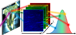
Nanoscale studies of bone provide key indicators to evidence subtle structural changes that may occur in the biomedical, forensic and archaeological contexts. One specific problem encountered in all those disciplines, for which the identification of nanostructural cues could prove useful, is to properly monitor the effect of heating on bone tissue. In particular, the mechanisms at work at the onset of heating are still relatively unclear. Using a multiscale approach combining Raman microspectroscopy, transmission electron microscopy (TEM), synchrotron quantitative scanning small-angle X-ray scattering imaging (qsSAXSI) and polarized light (PL) microscopy, we investigate the ultrastructure of cortical bovine bone heated at temperatures < 300 ̊C, from the molecular to the macroscopic scale. We show that, despite limited changes in crystal structure, the mineral nanoparticles increase in thickness and become strongly disorganized upon heating. Furthermore, while the nanostructure in distinct anatomical quadrants appears to be statistically different, our results demonstrate this stems from the tissue histology, i.e. from the high degree of heterogeneity of the microstructure induced by the complex cellular processes involved in bone tissue formation. From this study, we conclude that the analysis of bone samples based on the structure and organization of the mineral nanocrystals requires performing measurements at the histological level, which is an advantageous feature of qsSAXSI. This is a critical aspect that extends to a much broader range of questions relating to nanoscale investigations of bone, which could also be extended to other classes of nanostructured heterogeneous materials.
@article{gourrier2017nanoscale, title = {Nanoscale modifications in the early heating stages of bone are heterogeneous at the microstructural scale}, author = {Gourrier, Aur{\'e}lien and Chadefaux, C{\'e}line and Lemaitre, Estelle and Bellot-Gurlet, Ludovic and Reynolds, Michael and Burghammer, Manfred and Plazanet, Marie and Boivin, Georges and Farlay, Delphine and Bunk, Oliver and others}, journal = {PloS one}, volume = {12}, number = {4}, pages = {e0176179}, year = {2017}, publisher = {Public Library of Science San Francisco, CA USA}, url = {https://doi.org/10.1371/journal.pone.0176179}, doi = {10.1371/journal.pone.0176179} } - SciRepLabel-free imaging of bone multiscale porosity and interfaces using third-harmonic generation microscopyRachel Genthial, Emmanuel Beaurepaire, Marie-Claire Schanne-Klein, and 8 more authorsScientific Reports, 2017
Interfaces provide the structural basis of essential bone functions. In the hierarchical structure of bone tissue, heterogeneities such as porosity or boundaries are found at scales ranging from nanometers to millimeters, all of which contributing to macroscopic properties. To date, however, the complexity or limitations of currently used imaging methods restrict our understanding of this functional integration. Here we address this issue using label-free third-harmonic generation (THG) microscopy. We find that the porous lacuno-canalicular network (LCN), revealing the geometry of osteocytes in the bone matrix, can be directly visualized in 3D with submicron precision over millimetric fields of view compatible with histology. THG also reveals interfaces delineating volumes formed at successive remodeling stages. Finally, we show that the structure of the LCN can be analyzed in relation with that of the extracellular matrix and larger-scale structures by simultaneously recording THG and second-harmonic generation (SHG) signals relating to the collagen organization.
@article{genthial2017label, title = {Label-free imaging of bone multiscale porosity and interfaces using third-harmonic generation microscopy}, author = {Genthial, Rachel and Beaurepaire, Emmanuel and Schanne-Klein, Marie-Claire and Peyrin, Fran{\c{c}}oise and Farlay, Delphine and Olivier, C{\'e}cile and Bala, Yohann and Boivin, Georges and Vial, Jean-Claude and D{\'e}barre, Delphine and others}, journal = {Scientific Reports}, volume = {7}, number = {1}, pages = {3419}, year = {2017}, publisher = {Nature Publishing Group}, url = {https://www.nature.com/articles/s41598-017-03548-5}, doi = {10.1038/s41598-017-03548-5} } - NewJPhysStructure and dynamics of multicellular assemblies measured by coherent light scatteringBenjamin Brunel, Carles Blanch, Aurélien Gourrier, and 6 more authorsNew Journal of Physics, 2017
Determining the structure and the internal dynamics of tissues is essential to understand their functional organization. Microscopy allows for monitoring positions and trajectories of every single cell. Those data are useful to extract statistical observables, such as intercellular distance, tissue symmetry and anisotropy, and cell motility. However, this procedure requires a large and supervised computational effort. In addition, due to the large cross-section of cells, the light scattering limits the use of microscopy to relatively thin samples. As an alternative approach, we propose to take advantage of light scattering and to analyze the dynamical diffraction pattern produced by a living tissue illuminated with coherent light. In this article, we illustrate with a few examples that supra-cellular structures produce an exploitable diffraction signal. From the diffraction signal, we deduce the mean distance between cells, the anisotropy of the supra-cellular organization and, from its fluctuations, the mean speed of moving cells. This easy to implement technique considerably reduces analysis time, allowing real time monitoring.
@article{brunel_structure_2017,
  title = {Structure and dynamics of multicellular assemblies measured by coherent light scattering},
  author = {Brunel, Benjamin and Blanch, Carles and Gourrier, Aurélien and Petrolli, Vanni and Delon, Antoine and Joanny, Jean-François and Carminati, Rémi and Pierrat, Romain and Cappello, Giovanni},
  volume = {19},
  number = {7},
  pages = {073033},
  year = {2017},
  journal = {New Journal of Physics},
  url = {https://iopscience.iop.org/article/10.1088/1367-2630/aa7b0f},
  doi = {10.1088/1367-2630/aa7b0f}
}
2016
- BookChaptDe l’analyse des ivoires archéologiques dorés à la synthèse archéo-inspirée des nanoparticules hybrides pour les applications biomédicalesIna Reiche, Aurélien Gourrier, and Jolanda SpadavecchiaRegards croisés: quand les sciences archéologiques rencontrent l’innovation, 2016
@article{reiche2016analyse, title = {De l’analyse des ivoires arch{\'e}ologiques dor{\'e}s {\`a} la synth{\`e}se arch{\'e}o-inspir{\'e}e des nanoparticules hybrides pour les applications biom{\'e}dicales}, author = {Reiche, Ina and Gourrier, Aur{\'e}lien and Spadavecchia, Jolanda}, journal = {Regards crois{\'e}s: quand les sciences arch{\'e}ologiques rencontrent l’innovation}, pages = {123}, year = {2016}, publisher = {Archives contemporaines} } - BookChaptInformative Potential of Multiscale Observations in Archaeological Biominerals Down to NanoscaleIna Reiche and Aurélien GourrierIn Nanoscience and Cultural Heritage, 2016
Humans have intentionally used biological materials such as bone, ivory and shells since prehistoric times due to their particular physical and chemical properties. The composite nature of biological materials at the nanoscale combined with an important structural hierarchy up to the macroscopic level is responsible for these exceptional properties. In this chapter we discuss the relation of the structural features of different biological materials within their archaeological and historical contexts along with their anthropological use and function. Amid the wealth of biological materials of archaeological interest, a special attention is paid to carbonate–based materials such as corals and shells and phosphate-based ones, including bone, teeth, ivory and antler. The structural features of these archaeobiominerals at different length scales down to the nanoscopic scale are highlighted in this chapter as they allow drawing conclusions on ancient working techniques, the provision and circulation of raw materials, anthropological heat processes, and, last but not least, on diagenetic changes and authentication purposes. The informative potential of observations of archaeological biological materials at different length scales is finally illustrated by some case studies.
@incollection{reiche2016informative, title = {Informative Potential of Multiscale Observations in Archaeological Biominerals Down to Nanoscale}, author = {Reiche, Ina and Gourrier, Aur{\'e}lien}, booktitle = {Nanoscience and Cultural Heritage}, pages = {75--122}, year = {2016}, publisher = {Springer}, url = {http://link.springer.com/10.2991/978-94-6239-198-7_4}, doi = {10.2991/978-94-6239-198-7_4} } - TopicsCurChemEmerging approaches in synchrotron studies of materials from cultural and natural history collectionsLoı̈c Bertrand, Sylvain Bernard, Federica Marone, and 5 more authorsTopics in current chemistry, 2016
Synchrotrons have provided significant methods and instruments to study ancient materials from cultural and natural heritages. New ways to visualise (surfacic or volumic) morphologies are developed on the basis of elemental, density and refraction contrasts. They now apply to a wide range of materials, from historic artefacts to paleontological specimens. The tunability of synchrotron beams owing to the high flux and high spectral resolution of photon sources is at the origin of the main chemical speciation capabilities of synchrotron-based techniques. Although, until recently, photon-based speciation was mainly applicable to inorganic materials, novel developments based, for instance, on STXM and deep UV photoluminescence bring new opportunities to study speciation in organic and hybrid materials, such as soaps and organometallics, at a submicrometric spatial resolution over large fields of view. Structural methods are also continuously improved and increasingly applied to hierarchically structured materials for which organisation results either from biological or manufacturing processes. High-definition (spectral) imaging appears as the main driving force of the current trend for new synchrotron techniques for research on cultural and natural heritage materials.
@article{bertrand2016emerging,
  title = {Emerging approaches in synchrotron studies of materials from cultural and natural history collections},
  author = {Bertrand, Lo{\"\i}c and Bernard, Sylvain and Marone, Federica and Thoury, Mathieu and Reiche, Ina and Gourrier, Aur{\'e}lien and Sciau, Philippe and Bergmann, Uwe},
  journal = {Topics in current chemistry},
  volume = {374},
  number = {1},
  pages = {7},
  year = {2016},
  publisher = {Springer International Publishing},
  url = {http://dx.doi.org/10.1007/s41061-015-0003-1},
  doi = {10.1007/s41061-015-0003-1}
}
2015
- BookChaptChapitre 3 L’os: morphologie, structure et composition chimiqueAurélien Gourrier and Ina Reiche2015
@misc{gourrier2015chapitre,
  title = {Chapitre 3 L'os: morphologie, structure et composition chimique},
  author = {Gourrier, Aur{\'e}lien and Reiche, Ina},
  year = {2015},
  url = {http://hal.univ-grenoble-alpes.fr/hal-01131757/document},
}
2014
- Pal3Early diagenesis of elephant tusk in marine environmentMarie Albéric, Aurélien Gourrier, Katharina Müller, and 4 more authorsPalaeogeography, Palaeoclimatology, Palaeoecology, 2014
Three elephant tusks recovered from a 18th century Dutch shipwreck in Brittany seawaters were analyzed to study early diagenetic changes of ivory in a marine environment. Micro-small angle X-ray scattering (μSAXS) analysis revealed sub-microscopic structural modifications of ivory (hydroxyapatite particle thickness, mineralized collagen fibers organization and orientation). Chemical compositional changes of the organic fraction (nitrogen and carbon loss) and of the mineral part (magnesium loss, carbonate, calcium, chlorine and strontium uptake) were investigated by non-invasive micro-Particle Induced X-ray Emission/Gamma-ray Emission/Rutherford and Elastic Backscattering Spectrometry analysis (μPIXE/PIGE/RBS/EBS). These chemical and structural markers can serve as proxies for the early diagenesis of elephant tusk in marine settings. The results show that the macroscopic preservation state of the tusk surface does not reflect its microscopic preservation state. A diagenetic sequence is proposed, depending on the local environment of each tusk. First, collagen hydrolysis and ion adsorptions from the marine environment are proposed to occur. Second, a tusk buried in the sand was protected from Ca enrichment of the surface and the formation of a flaky white concretion whereas tusks out of the sand were subjected to these latter phenomena as well as sea-urchin attacks. Third, ion leaching and uptake, increased crystallinity of the hydroxyapatite crystals, and the modifications of the mineralized collagen fiber network are assumed to happen for all three tusks with different level of completion.
@article{alberic2014early,
  title = {Early diagenesis of elephant tusk in marine environment},
  author = {Alb{\'e}ric, Marie and Gourrier, Aur{\'e}lien and M{\"u}ller, Katharina and Zizak, I and Wagermaier, Wolfgang and Fratzl, Peter and Reiche, Ina},
  journal = {Palaeogeography, Palaeoclimatology, Palaeoecology},
  volume = {416},
  pages = {120--132},
  year = {2014},
  publisher = {Elsevier},
  url = {http://dx.doi.org/10.1016/j.palaeo.2014.09.006},
  doi = {10.1016/j.palaeo.2014.09.006}
}
2013
- BookChaptUnderstanding Hierarchy and Functions of Bone Using Scanning X‐ray Scattering MethodsWolfgang Wagermaier, Aurélien Gourrier, and Barbara AichmayerIn Smart Materials Series, 2013
@incollection{wagermaier2013understanding, title = {Understanding Hierarchy and Functions of Bone Using Scanning X‐ray Scattering Methods}, booktitle = {Smart Materials Series}, author = {Wagermaier, Wolfgang and Gourrier, Aur{\'e}lien and Aichmayer, Barbara}, editor = {Fratzl, Peter and Dunlop, John W.C. and Weinkamer, Richard}, pages = {46--73}, year = {2013}, publisher = {Royal Society of Chemistry}, url = {https://doi.org/10.1039/9781849737555-00046}, doi = {10.1039/9781849737555-00046} } - PLOSOneMicrofibril orientation dominates the microelastic properties of human bone tissue at the lamellar length scaleMathilde Granke, Aurélien Gourrier, Fabienne Rupin, and 5 more authorsPLoS One, 2013
The elastic properties of bone tissue determine the biomechanical behavior of bone at the organ level. It is now widely accepted that the nanoscale structure of bone plays an important role to determine the elastic properties at the tissue level. Hence, in addition to the mineral density, the structure and organization of the mineral nanoparticles and of the collagen microfibrils appear as potential key factors governing the elasticity. Many studies exist on the role of the organization of collagen microfibril and mineral nanocrystals in strongly remodeled bone. However, there is no direct experimental proof to support the theoretical calculations. Here, we provide such evidence through a novel approach combining several high resolution imaging techniques: scanning acoustic microscopy, quantitative scanning small-Angle X-ray scattering imaging and synchrotron radiation computed microtomography. We find that the periodic modulations of elasticity across osteonal bone are essentially determined by the orientation of the mineral nanoparticles and to a lesser extent only by the particle size and density. Based on the strong correlation between the orientation of the mineral nanoparticles and the collagen molecules, we conclude that the microfibril orientation is the main determinant of the observed undulations of microelastic properties in regions of constant mineralization in osteonal lamellar bone. This multimodal approach could be applied to a much broader range of fibrous biological materials for the purpose of biomimetic technologies.
@article{granke2013microfibril,
  title = {Microfibril orientation dominates the microelastic properties of human bone tissue at the lamellar length scale},
  author = {Granke, Mathilde and Gourrier, Aur{\'e}lien and Rupin, Fabienne and Raum, Kay and Peyrin, Fran{\c{c}}oise and Burghammer, Manfred and Sa{\"\i}ed, Amena and Laugier, Pascal},
  journal = {PLoS One},
  volume = {8},
  number = {3},
  pages = {e58043},
  year = {2013},
  publisher = {Public Library of Science},
  url = {http://dx.doi.org/10.1371/journal.pone.0058043},
  doi = {10.1371/journal.pone.0058043}
}
2012
- BBNSynchrotron 3D SAXS analysis of bone nanostructureRobin Seidel, Aurelien Gourrier, Michael Kerschnitzki, and 4 more authorsBioinspired, Biomimetic and Nanobiomaterials, 2012
The complex structure of bone requires a structural description of the material at different hierarchical levels. At the micrometer level, collagen fibril orientation and osteocyte network architecture can be described by different light microscopy methods. However, further investigation of the nanostructure of bone requires high-resolution techniques such as electron microscopy as well as x-ray scattering methods. The basic building blocks at the nanometer level are organic type I collagen fibrils reinforced by nanoparticles of carbonated apatite mineral. Most commonly, these fibrils aggregate into lamellae of about 5 µm width, in both compact and spongy bone. The architecture of the mineral platelets and the collagen fibrils influences the mechanical properties. Models of twisted and rotated plywood motifs have been proposed, although detailed quantitative characterization at length scales comparable to typical tissue unit sizes are still lacking. Here, we describe a scanning small-angle x-ray scattering (SAXS) method to reconstruct the variation of the three-dimensional habit of mineral platelets within osteonal bone. We find that the platelets change their orientation at micrometer resolution and are organized structurally by a repeating unit of about 5 µm, which is in agreement with previous wide-angle x-ray diffraction microtexture measurements. At the spatial resolution of the microbeam used (1 µm), we observe fiber geometry. The presented SAXS reconstruction technique could also be applied to the analysis of nanoparticle orientation in highly textured biomaterials.
- PNASStructure-property relationships of a biological mesocrystal in the adult sea urchin spineJong Seto, Yurong Ma, Sean A Davis, and 8 more authorsProceedings of the National Academy of Sciences, 2012
Structuring over many length scales is a design strategy widely used in Nature to create materials with unique functional properties. We here present a comprehensive analysis of an adult sea urchin spine, and in revealing a complex, hierarchical structure, show how Nature fabricates a material which diffracts as a single crystal of calcite and yet fractures as a glassy material. Each spine comprises a highly oriented array of Mg-calcite nanocrystals in which amorphous regions and macromolecules are embedded. It is postulated that this mesocrystalline structure forms via the crystallization of a dense array of amorphous calcium carbonate (ACC) precursor particles. A residual surface layer of ACC and/or macromolecules remains around the nanoparticle units which creates the mesocrystal structure and contributes to the conchoidal fracture behavior. Nature’s demonstration of how crystallization of an amorphous precursor phase can create a crystalline material with remarkable properties therefore provides inspiration for a novel approach to the design and synthesis of synthetic composite materials.
@article{seto2012structure,
  title = {Structure-property relationships of a biological mesocrystal in the adult sea urchin spine},
  author = {Seto, Jong and Ma, Yurong and Davis, Sean A and Meldrum, Fiona and Gourrier, Aurelien and Kim, Yi-Yeoun and Schilde, Uwe and Sztucki, Michael and Burghammer, Manfred and Maltsev, Sergey and others},
  journal = {Proceedings of the National Academy of Sciences},
  volume = {109},
  number = {10},
  pages = {3699--3704},
  year = {2012},
  publisher = {National Acad Sciences},
  url = {https://pnas.org/doi/full/10.1073/pnas.1109243109},
  doi = {10.1073/pnas.1109243109}
}
2011
- ConfAbstractModeling the organization of molecules in collagen using the paracrystal conceptJean Doucet, Fatma Briki, Aurelien Gourrier, and 4 more authorsActa Crystallographica Section A, 2011
- ConfProcArtificially heated bone at low temperatures: a quantitative scanning-small-angle X-ray scattering imaging study of the mineral particle sizeAurélien Gourrier, O Bunk, Katharina Müller, and 1 more authorArcheoSciences. Revue d’archéométrie, 2011
The ultrastructure of bovine femoral bone artificially heated at low temperatures (\textless 300 °C) is investigated by means of synchrotron quantitative scanning-SAXS imaging. Significant changes in the distribution of particle size are observed upon heating. On average, an exponential particle growth is measured with increasing temperature independently of the tissue histology. Additionally, the heating process induces a broader dispersion in the particle size which, in turn, seems to be dependent on the bone microstructure. Those parameters could therefore be used as markers in the characterization of archaeological bone presenting traces of heating.
- ConfProcTowards a better understanding of alteration phenomena of archaeological bone by a closer look at the organic/mineral association at micro-and nanoscale. Preliminary results on Neolithic samples from Chalain lake site 19, Jura, FranceIna Reiche, Céline Chadefaux, Katharina Müller, and 1 more authorArcheoSciences. Revue d’archéométrie, 2011
In this paper we present the extension of existing analytical schemes for the precise evaluation of archaeological bone preservation states. The new methodological developments concerning the study of the morphological and structural features of archaeological bones at micro- and nanoscale are emphasized in order to elucidate fine diagenetic modifications and to better understand the underlying alteration mechanisms. A combination of synchrotron X-ray microtomography, infrared micro-spectroscopy imaging and quantitative scanning small-angle X-ray scattering (sSAXS) imaging allowed studying micromorphological changes and the distribution of the organic and mineral components of bone at a histological level. Transmission electron microscopy gives precise information on changes of apatite crystal dimensions, orientations, periodicity of collagen fiber arrangement and crystal distributions within the fibers in archaeological bone. It allows establishing criteria to evaluate more precisely the preservation state of archaeological bone material with respect to modern references. The potential of these methods is highlighted on the basis of the study of some macroscopically well preserved archaeological bone samples from the Chalain lake station 19, Neolithic period, Jura, France. The investigations allowed a differentiation in preservation state between these samples and the postulation of an alteration sequence in such humid chalk-rich burial environments.
- ConfAbstractPulmonary Lesions Among Welders Exposed To Nanoparticles From Welding FumesPascal Andujar, Angélique Simon-Deckers, Barbara Fayard, and 8 more authorsIn A96. NANOPARTICLES, 2011
- JSBModeling the lateral organization of collagen molecules in fibrils using the paracrystal conceptJean Doucet, Fatma Briki, Aurélien Gourrier, and 4 more authorsJournal of structural biology, 2011
A characteristic feature of the dense phases formed by fiber-shaped molecules is their organization into parallel rods packed in a hexagonal or pseudo-hexagonal lateral network. This is typically the case for the collagen triple helices inside fibrils, as confirmed by recent X-ray diffraction experiments carried out on highly crystallized fibers obtained by immersing the freshly extracted fibers in a salt-controlled medium. However such diffraction patterns also generally exhibit additional features in the form of diffuse scattering, which is a clear signature of a low degree of lateral ordering. Only few studies have analyzed and modeled the lateral packing of collagen triple helices when the structure is disordered. Some authors have used the concept of short-range order but this approach does not contain any echo of a hexagonal order. In this study, we use an analytical expression derived from the paracrystal model which retains the hexagonal symmetry information and leads to a good agreement with the experimental data in the medium-angle region. This method is quite sensitive to the degree of disorder and to the inter-object distance. One clear result is that the shift in peak positions, generally attributed to variations in intermolecular distances, can also arise from a change in the degree of ordering without any significant modification of the distances. This underlines the importance of evaluating the degree of ordering before attributing a shift in peak position to a change in the unit-cell. This method is generic and can be applied to any system composed of rod-shaped molecules.
@article{doucet2011modelinh,
  title = {Modeling the lateral organization of collagen molecules in fibrils using the paracrystal concept},
  author = {Doucet, Jean and Briki, Fatma and Gourrier, Aur{\'e}lien and Pichon, Chantal and Gumez, Laurie and Bensamoun, Sabine and Sadoc, Jean-Fran{\c{c}}ois},
  journal = {Journal of structural biology},
  volume = {173},
  number = {2},
  pages = {197--201},
  year = {2011},
  publisher = {Elsevier},
  url = {http://dx.doi.org/10.1016/j.jsb.2010.11.018},
  doi = {10.1016/j.jsb.2010.11.018}
}
2010
- ConfAbstractRecent developments in scanning micro and nanobeam diffraction techniquesManfred Burghammer, Sebastian Schöder, Silvia Santucci, and 4 more authorsActa Crystallographica Section A, 2010
- JSBHard alpha-keratin degradation inside a tissue under high flux X-ray synchrotron micro-beam: A multi-scale time-resolved studyEmilie Leccia, Aurélien Gourrier, Jean Doucet, and 1 more authorJournal of structural biology, 2010
X-rays interact strongly with biological organisms. Synchrotron radiation sources deliver very intense Xray photon fluxes within micro- or submicro cross-section beams, resulting in doses larger than the MGy. The relevance of synchrotron radiation analyses of biological materials is therefore questionable since such doses, million times higher than the ones used in radiotherapy, can cause huge damages in tissues, with regard to not only DNA, but also proteic and lipid organizations. Very few data concerning the effect of very high X-ray doses in tissues are available in the literature. We present here an analysis of the structural phenomena which occur when the model tissue of human hair is irradiated by a synchrotron X-ray micro-beam. The choice of hair is supported by its hierarchical and partially ordered keratin structure which can be analysed inside the tissue by X-ray diffraction. To assess the damages caused by hard Xray micro-beams (1 lm2 cross-section), short exposure time scattering SAXS/WAXS patterns have been recorded at beamline ID13 (ESRF) after various irradiation times. Various modifications of the scattering patterns are observed, they provide fine insight of the radiation damages at various hierarchical levels and also unexpectedly provide information about the stability of the various hierarchical structural levels. It appears that the molecular level, i.e. the alpha helices which are stabilized by hydrogen bonds and the alpha-helical coiled coils which are stabilized by hydrophobic interactions, is more sensitive to radiation than the supramolecular architecture of the keratin filament and the filament packing within the keratin associated proteins matrix, which is stabilized by disulphide bonds.
@article{leccia2010hard, title = {Hard alpha-keratin degradation inside a tissue under high flux X-ray synchrotron micro-beam: A multi-scale time-resolved study}, author = {Leccia, Emilie and Gourrier, Aur{\'e}lien and Doucet, Jean and Briki, Fatma}, journal = {Journal of structural biology}, volume = {170}, number = {1}, pages = {69--75}, year = {2010}, publisher = {Elsevier}, url = {http://dx.doi.org/10.1016/j.jsb.2009.11.006}, doi = {10.1016/j.jsb.2009.11.006} } - JACScanning small-angle X-ray scattering analysis of the size and organization of the mineral nanoparticles in fluorotic bone using a stack of cards modelAurélien Gourrier, Chenghao Li, Stefan Siegel, and 4 more authorsJournal of Applied Crystallography, 2010
A model describing the size and arrangement of mineral particles in bone tissues is used to analyse the results of a scanning small-angle X-ray scattering (SAXS) experiment on a pathological bone biopsy. The overall description assumes that the nanometre-sized mineral platelets are arranged in a parallel fashion with possible fluctuations in their relative position, orientation and thickness. This method is tested on a thin sample section obtained from the biopsy of an osteoporotic patient treated with a high cumulative dose of NaF. The mineralization pattern of fluorotic bone is known to exhibit significant differences as compared to healthy bone in terms of density, particle size and organization. This is the first attempt to provide quantitative indicators of the degree of regularity in the packing of the mineral platelets in human pathological bone. Using scanning SAXS with a synchrotron microbeam of 15µm allows discrimination between pathological and healthy bone at the tissue level. Additionally, the benefits of this method are discussed with respect to the accuracy of particle size determination using SAXS.
@article{gourrier2010scanning,
  title = {Scanning small-angle X-ray scattering analysis of the size and organization of the mineral nanoparticles in fluorotic bone using a stack of cards model},
  author = {Gourrier, Aur{\'e}lien and Li, Chenghao and Siegel, Stefan and Paris, Oskar and Roschger, Paul and Klaushofer, Klaus and Fratzl, Peter},
  journal = {Journal of Applied Crystallography},
  volume = {43},
  number = {6},
  pages = {1385--1392},
  year = {2010},
  publisher = {International Union of Crystallography},
  url = {http://dx.doi.org/10.1107/S0021889810035090},
  doi = {10.1107/S0021889810035090}
}
2009
- CalcifTissueIntCombination of nanoindentation and quantitative backscattered electron imaging revealed altered bone material properties associated with femoral neck fragilityN Fratzl-Zelman, P Roschger, A Gourrier, and 6 more authorsCalcified tissue international, 2009
Osteoporotic fragility fractures were hypothesized to be related to changes in bone material properties and not solely to reduction in bone mass. We studied cortical bone from the superior and inferior sectors of whole femoral neck sections from five female osteoporotic hip fracture cases (74–92 years) and five nonfractured controls (75–88 years). The typical calcium content (CaPeak) and the mineral particle thickness parameter (T) were mapped in large areas of the superior and inferior regions using quantitative backscattered electron imaging (qBEI) and scanning small-angle X-ray scattering, respectively. Additionally, indentation modulus (E) and hardness (H) (determined by nanoindentation) were compared at the local level to the mineral content (CaInd) at the indent positions (obtained from qBEI). CaPeak (-2.2%, P = 0.002), CaInd (-1.8%, P = 0.048), E (-5.6%, P = 0.040), and H (-6.0%, P = 0.016) were significantly lower for the superior compared to the inferior region. Interestingly, CaPeak as well as CaInd were also lower (-2.6%, P = 0.006, and –3.7%, P = 0.002, respectively) in fracture cases compared to controls, while E and H did not show any significant reduction. T values were in the normal range, independent of region (P = 0.181) or fracture status (P = 0.551). In conclusion, it appears that the observed femoral neck fragility is associated with a reduced mineral content, which was not accompanied by a reduction in stiffness and hardness of the bone material. This pilot study suggests that a stiffening process in the organic matrix component contributes to bone fragility independently of mineral content.
@article{fratzl2009combination,
  title = {Combination of nanoindentation and quantitative backscattered electron imaging revealed altered bone material properties associated with femoral neck fragility},
  author = {Fratzl-Zelman, N and Roschger, P and Gourrier, A and Weber, M and Misof, BM and Loveridge, N and Reeve, J and Klaushofer, K and Fratzl, P},
  journal = {Calcified tissue international},
  volume = {85},
  number = {4},
  pages = {335--343},
  year = {2009},
  publisher = {Springer-Verlag New York},
  url = {http://dx.doi.org/10.1007/s00223-009-9289-8},
  doi = {10.1007/s00223-009-9289-8}
}
2008
- MicronMapping fibre orientation in complex-shaped biological systems with micrometre resolution by scanning X-ray microdiffractionRobin Seidel, Aurélien Gourrier, Manfred Burghammer, and 3 more authorsMicron, 2008
A fully automated procedure to extract and to image local fibre orientation in biological tissues from scanning X-ray diffraction is presented. The preferred chitin fibre orientation in the flow sensing system of crickets is determined with high spatial resolution by applying synchrotron radiation based X-ray microbeam diffraction in conjunction with advanced sample sectioning using a UV micro-laser. The data analysis is based on an automated detection of azimuthal diffraction maxima after 2D convolution filtering (smoothing) of the 2D diffraction patterns. Under the assumption of crystallographic fibre symmetry around the morphological fibre axis, the evaluation method allows mapping the three-dimensional orientation of the fibre axes in space. The resulting two-dimensional maps of the local fibre orientations – together with the complex shape of the flow sensing system – may be useful for a better understanding of the mechanical optimization of such tissues.
@article{seidel2008mapping,
  title = {Mapping fibre orientation in complex-shaped biological systems with micrometre resolution by scanning X-ray microdiffraction},
  author = {Seidel, Robin and Gourrier, Aur{\'e}lien and Burghammer, Manfred and Riekel, Christian and Jeronimidis, George and Paris, Oskar},
  journal = {Micron},
  volume = {39},
  number = {2},
  pages = {198--205},
  year = {2008},
  publisher = {Elsevier},
  url = {http://dx.doi.org/10.1016/j.micron.2007.02.004},
  doi = {10.1016/j.micron.2007.02.004}
}
2007
- JACScanning X-ray imaging with small-angle scattering contrastAurelien Gourrier, Wolfgang Wagermaier, Manfred Burghammer, and 6 more authorsJournal of Applied Crystallography, 2007
- JACScanning texture analysis of lamellar bone using microbeam synchrotron X-ray radiationWolfgang Wagermaier, Himadri S Gupta, Aurélien Gourrier, and 5 more authorsJournal of Applied Crystallography, 2007
Texture analysis with microbeam scanning diffraction enables the local mapping of three-dimensional crystallite orientation in heterogeneous natural and synthetic materials. Cortical (compact) bone is an example of a hierarchically structured biocomposite, which is built mainly of cylindrical osteons, having a lamellar texture at the micrometre level. In this work, a combination of microbeam synchrotron X-ray texture analysis with thin sections of osteonal bone is used to measure the three-dimensional distribution of the c-axis orientation of the mineral apatite in bone with positional resolution of 1µm. The data reduction procedure needed to go from the stereographic projection of X-ray intensity to the determination of the local orientation of mineralized collagen fibrils is described. The procedure can be applied to other mineralized tissues (such as trabecular bone and chitin) with micrometre scale and biologically controlled fibrillar texture.
@article{wagermaier2007scanning,
  title = {Scanning texture analysis of lamellar bone using microbeam synchrotron X-ray radiation},
  author = {Wagermaier, Wolfgang and Gupta, Himadri S and Gourrier, Aur{\'e}lien and Paris, Oskar and Roschger, Paul and Burghammer, Manfred and Riekel, Christian and Fratzl, Peter},
  journal = {Journal of Applied Crystallography},
  volume = {40},
  number = {1},
  pages = {115--120},
  year = {2007},
  publisher = {International Union of Crystallography},
  url = {http://dx.doi.org/10.1107/S0021889806044888},
  doi = {10.1107/S0021889806044888}
}
2006
- PhilMagHardness testing under a different light: combining synchrotron X-ray microdiffraction and indentation techniques for polymer fibres studiesAurélien Gourrier, MC Garcı́a-Gutiérrez, and Christian RiekelPhilosophical Magazine, 2006
- BiointerphSpiral twisting of fiber orientation inside bone lamellaeW Wagermaier, H S. Gupta, A Gourrier, and 3 more authorsBiointerphases, 2006
The secondary osteon — a fundamental building block in compact bone — is a multilayered cylindrical structure of mineralized collagen fibrils arranged around a blood vessel. Functionally, the osteon must be adapted to the in vivo mechanical stresses in bone at the level of its microstructure. However, questions remain about the precise mechanism by which this is achieved. By application of scanning x-ray diffraction with a micron-sized synchrotron beam, along with measurements of local mineral crystallographic axis direction, we reconstruct the three-dimensional orientation of the mineralized fibrils within a single osteon lamella (∼5 μm). We find that the mineralized collagen fibrils spiral around the central axis with varying degrees of tilt, which would — structurally — impart high extensibility to the osteon. As a consequence, strains inside the osteon would have to be taken up by means of shear between the fibrils.
@article{wagermaier2006spiral,
  title = {Spiral twisting of fiber orientation inside bone lamellae},
  author = {Wagermaier, W and S. Gupta, H and Gourrier, A and Burghammer, M and Roschger, P and Fratzl, P},
  journal = {Biointerphases},
  volume = {1},
  number = {1},
  pages = {1--5},
  year = {2006},
  publisher = {AVS},
  url = {https://pubs.aip.org/bip/article/1/1/1/124079/Spiral-twisting-of-fiber-orientation-inside-bone},
  doi = {10.1116/1.2178386}
}
2005
- ConfAbstractMicrobeam Diffraction of Hierarchical NanocompositesOskar Paris, Peter Fratzl, Aurelien Gourrier, and 6 more authorsACTA CRYSTALLOGRAPHICA A-FOUNDATION AND ADVANCES, 2005
- MacromolInvestigation of the structural deformation mechanisms induced by microindentation in a thermotropic liquid crystalline copolyester using synchrotron x-ray microdiffractionAurélien Gourrier, Mari-Cruz Garcı́a Gutiérrez, and Christian RiekelMacromolecules, 2005
The structural changes induced by microindentation on thermotropic liquid crystalline copolyester fibers of 4-hydroxybenzoic acid (HBA) and 2-hydroxy-6-naphthoic acid (HNA) have been investigated by scanning X-ray microdiffraction techniques at the ESRF microfocus beamline (ID13). A phase transformation is observed upon deformation from a pseudohexagonal lattice (PH) to two distinct orthorhombic phases (OI, OII). In addition, a strong texture is observed in the plastically deformed region as revealed by changes in the local orientation of the crystalline domains in the vicinity of the indenter tip and the appearance of reflections in specific directions only. This allows quantifying the extent of the deformation both across and along the fiber and also suggests the presence of complex structural processes occurring during indentation tests.
@article{gourrier2005investigation,
  title = {Investigation of the structural deformation mechanisms induced by microindentation in a thermotropic liquid crystalline copolyester using synchrotron x-ray microdiffraction},
  author = {Gourrier, Aur{\'e}lien and Garc{\'\i}a Guti{\'e}rrez, Mari-Cruz and Riekel, Christian},
  journal = {Macromolecules},
  volume = {38},
  number = {9},
  pages = {3838--3844},
  year = {2005},
  publisher = {ACS Publications},
  url = {http://dx.doi.org/10.1021/ma047509p},
  doi = {10.1021/ma047509p}
}
2004
- JMacromolSciBInvestigation of structural processes during indentation of polymers by synchrotron radiation microdiffractionMC Garcı́a Gutiérrez, A Gourrier, and C RiekelJournal of Macromolecular Science, Part B, 2004
A Vickers microindentation setup has been integrated into the scanning setup used at the European Synchrotron Radiation Facility (ESRF) microfocus beamline (ID 13). Ex situ wide-angle x-ray scattering experiments performed on an isotactic polypropylene (iPP) single fiber show changes in chain orientation and breakup of the monoclinic a-modification into domains due to plastic deformation in the indented zone. The systematic mapping of the perturbed zone allows spatially dependant structural changes to be determined. Similar experiments for a Vectra single fiber show a partial transformation of the pseudohexagonal phase into the orthorhombic phase. In situ experiments allow following structural changes as a function of time and applied force. During indentation of an iPP fiber, domain doubling during indentation and single domain recovery after the tip has been retracted is observed.
2003
- AbalBioanalChemRecent synchrotron radiation microdiffraction experiments on polymer and biopolymer fibersC Riekel, MC Garcı́a Gutiérrez, A Gourrier, and 1 more authorAnalytical and bioanalytical chemistry, 2003
The status of synchrotron radiation (SR) microdiffraction techniques developed at the ID13 beamline of the European Synchrotron Radiation Facility (ESRF) is reviewed for polymer and biopolymer fiber applications. Beam sizes in the micrometer-range have been used to study the local structure of whole fibers such as viscoserayon or poly(p-phenylene terephthalamide). The possibilities for in situ studies during stretching, extrusion, or indentation will be discussed.
@article{riekel2003recent,
  title = {Recent synchrotron radiation microdiffraction experiments on polymer and biopolymer fibers},
  author = {Riekel, C and Guti{\'e}rrez, MC Garc{\'\i}a and Gourrier, A and Roth, S},
  journal = {Analytical and bioanalytical chemistry},
  volume = {376},
  number = {5},
  pages = {594--601},
  year = {2003},
  publisher = {Springer},
  doi = {10.1007/s00216-003-1976-0},
  url = {http://dx.doi.org/10.1007/s00216-003-1976-0},
}
2002
- MacromolCombined microindentation and synchrotron radiation microdiffraction applied to polymersA Gourrier, MC Garcı́a Gutiérrez, and C RiekelMacromolecules, 2002
A Vickers microindentation setup has been developed for in situ X-ray microdiffraction with synchrotron radiation at the ESRF microfocus beamline (ID13). Ex situ experiments performed on a Nylon 66 fiber show changes in chain orientation in the indented zone, but the crystallinity remains unchanged. Similar experiments for an UHMW-PE fiber show a breakup of the orthorhombic phase into domains due to plastic deformation and a partial transformation of the orthorhombic phase into the monoclinic phase. In situ experiments during indentation of an UHMW-PE fiber show domain doubling during indentation and single domain recovery after the tip has been retracted.
@article{gourrier2002combined,
  title = {Combined microindentation and synchrotron radiation microdiffraction applied to polymers},
  author = {Gourrier, A and Garc{\'\i}a Guti{\'e}rrez, MC and Riekel, C},
  journal = {Macromolecules},
  volume = {35},
  number = {21},
  pages = {8072--8077},
  year = {2002},
  publisher = {ACS Publications},
  url = {http://dx.doi.org/10.1021/ma0206725},
  doi = {10.1021/ma0206725}
}
2000
- JSSCSynthesis, Structure, and Magnetic Behavior of La5Re3MnO16: A New Perovskite-like MaterialC.R. Wiebe, A. Gourrier, T. Langet, and 2 more authorsJournal of Solid State Chemistry, 2000
@article{wiebe_synthesis_2000,
  title = {Synthesis, Structure, and Magnetic Behavior of La5Re3MnO16: A New Perovskite-like Material},
  author = {Wiebe, C.R. and Gourrier, A. and Langet, T. and Britten, J.F. and Greedan, J.E.},
  journal = {Journal of Solid State Chemistry},
  volume = {151},
  number = {1},
  pages = {31--39},
  year = {2000},
  url = {http://dx.doi.org/10.1006/jssc.1999.8618},
  doi = {10.1006/jssc.1999.8618}
}