Tooth Dentin Porosity
Characterize the cellular porosity network and its functional impact
This project started in 2012 in collaboration with Elsa Vennat from CentraleSupélec, University Paris-Saclay, France.
Keywords: Tooth dentin, cellular porosity, odontoblasts, sentivity, biomechanics.
Our goal is to characterize dentinal porosity at the whole tooth level and understand its biomechanical impact and role in biomechanics and teeth sensitivy.
Origin of the project This project started with the observation of dentinal porosity close to the dentin-enamel junction (DEJ) using confocal fluorescence microscopy. Visualizing a dense 3D distribution of fine branches stemming out from the main tubules and performing finite element modeling (FEM) on idealized porosity representations, we got a first hint that dentinal porosity could clearly affect mechanical properties, with effects mittigated by the presence of a pertitubular collar (Vennat et al., 2017). In doing so, we realized that there was only limited knowledge about the porosity topology and connectivity, and even less about its precise functional role.
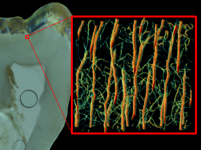
Optimizing optical fluorescence microscopy of dentinal porosity The first step to characterizing dentinal porosity is to be able to obtain statistically accurate 3D visalizations. This is the current objectif of SeungHwan Lee’s PhD.
The use of autofluorescence arrizing from endogenous fluorophores is another way to futher characterize dentin tissue. It allows overcoming situations where the porosity cannot be stained with a dye, e.g. when occluded by mineral, which frequently happens with ageing. In addition, it allows visualizing structural features that would require specific staining or complementary correlated measurements. A complete account can be found in: (Lee et al., 2022).
Characterization of the porosity network While tubules radiating from the pulp to the enamel are well-known porosity features, the distribution of secondary branches is much less clear from the littearture. During his PhD, Lucas Chatelain used graph analysis to show how those secondary branches mostly connect neighbouring tubules up to 1 mm away from the DEJ, thus forming a densely interconnected network (Chatelain et al., 2025). Following an in-depth quantification of errors, he also demonstrated that network characteristics strongly fluctuate in space, which may provide valuable histological indications of specific dentinogenesis events in healthy or pathological states.
Towards dentinal porosity imaging at the whole tooth level The studies perform so far clearly show that there is a strong need for extended imaging of dentinal porosity at the tooth level to be able to understand global function and possible pathological alterations. This is goal is currently persued by Lauren Anderson’s PhD.